Limbal Stem Cell Deficiency & Eye Prosthesis
April 7, 2016
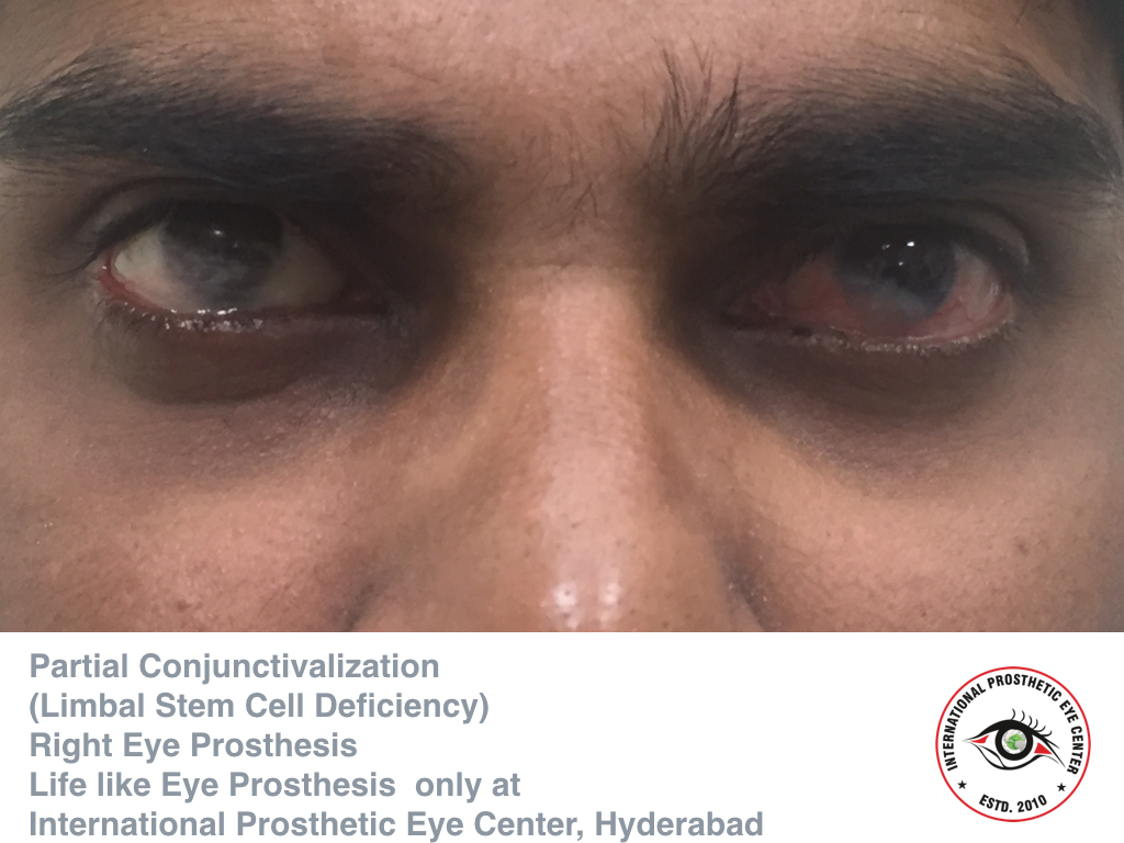
The corneal epithelium is a stratified squamous epithelium from which superficial terminal cells are naturally shed. Limbal stem cell deficiency (LSCD) is characterized by a loss or deficiency of the stem cells in the limbus that are vital for re-population of the corneal epithelium and to the barrier function of the limbus [1 2]. When these stem cells are lost, the corneal epithelium is unable to repair and renew itself. This results in epithelial breakdown and persistent epithelial defects, corneal conjunctivalization and neovascularization, corneal scarring, and chronic inflammation. All of these contribute to loss of corneal clarity, potential vision loss, chronic pain, photophobia, and keratoplasty failure[2 3].
Etiologies:
The etiologies can be genetic, acquired, or idiopathic.
Genetic:
Limbal stem cell deficiency has been associated with PAX6 gene mutations, which are also implicated in aniridia[4] and Peter’s Anomaly[5]. Other genetic disorders that have been reported with LSCD include ectrodactylyl-ectodermal-dysplasia-clefting syndrome[6], keratitis-ichthyosis-deafness (KID) Syndrome[7], Xeroderma Pigmentosum[8], Dominantly Inherited Keratitis[9], Turner Syndrome[3] and Dyskeratosis Congenita[10].
Acquired:
Inflammatory:
Other causes include inflammatory insults such as those seen in Steven-Johnsons Syndrome (SJS) [11], ocular cicatricial pemphigoid[12], and graft versus host disease[13]. Chronic ocular allergy such as Vernal Keratoconjunctivitis is another reported cause[14]. Neurotrophic keratopathy, whether neuronal or ischemic, can lead to this disease as well[2], as can bullous keratopathy [15].
Infectious:
Any infections of the corneal surface such as herpes keratitis[16] and trachoma [17] can predispose to this condition.
Traumatic/Iatrogenic:
Acquired causes also include trauma from chemical or thermal burns, and patients who have undergone prior ocular surgeries or cryotherapies at the limbus may be more susceptible[16 18]. Radiation and chemotherapy are other potential causes, and systemic[19] as well as topical chemotherapeutic medications may be sufficient to cause deficiency[20]. LSCD has also been seen with benzalkonium chloride toxicity with glaucoma medications [21]. Inappropriate contact lens use with consequent hypoxia and ocular irritation with destruction of the limbus may also contribute to both focal and total limbal stem cell deficiency[22 23].
Tumors/Overgrowth of Other Tissue:
Ocular surface tumors are a known cause of LSCD[2]. Pterygium may also cause a focal acquired absence of limbal stem cells[24].
Management:
Management is typically symptomatic in nature early in the disease. When limbal stem cell injury is transient, sometimes termed limbal stem cell disease or limbal stem cell distress, conservative medical measures as above may be sufficient[21 31 38] However, total limbal stem cell deficiency must be surgically managed.
Complication:
Untreated limbal stem cell deficiency causes pain, decreased vision, and recurrent epithelial erosions that predispose to infection and loss of vision. Infectious keratitis is common with this disease, and patients who wear contact lenses for extended periods of time, have persistent epithelial defects, and use topical immunosuppressive medications are at increased risk[32]. After surgical treatment, there is a risk of rejection from allogeneic transplants[49]. It is possible that the cornea will not remain clear and further surgery such as repeat stem cell transplant or penetrating keratoplasty may be necessary[49]
Cultivated Oral Mucosal Epithelial Transplantation (COMET):
Patients with live related stem cell transplantation or cultivated oral mucosal epithelial transplantation (COMET) along with lamellar or penetrating keratoplasty have poor outcomes even with long-term immunosuppression[54-56]. The use of fibrin glue rather than amniotic membrane for COMET and optimizing the ocular surface prior to transplant improved outcomes in a recent study, and it is possible that future modifications to technique may improve these outcomes further[57].
Cultivated Limbal Epithelial Transplantation (CLET):
Studies have shown that CLET is as effective as direct limbal transplantation for LSCD while requiring less donor tissue and thus being safer for donor eyes[45 58-61]. Studies of CLET have shown a 68-80% success rate.[62 63] In a review of outcomes of cultured limbal epithelial cell therapy published from 1997 to 2011 with data from 583 patients, the overall success rate was 76%[60]. However, this varies by The success rate of a transplant is significantly higher with an increased number of transplanted stem cells and failures tend to happen within the first year[63].
The largest study of xeno-free explant culture transplants showed a 71% success rate in 200 recipient eyes with a mean follow-up of approximately 5 years and up to 10 years.[46 49] Supplemental corneal transplant (PK) has a survival rate of 1 year, with a median survival of 3.3 years.[49]
In a recent meta-analysis of the outcomes of keratolimbal allografting for LSCD, postoperative corrected distance visual acuity (CDVA) was 2 or more lines better than the preoperative visual acuity in 31%to 67% of eyes [55].
Simple Limbal Epithelial Transplant:
In a study of 6 patients with total unilateral LSCD, visual acuity improved from worse than 20/200 in all recipient eyes before SLET surgery to 20/60 or better in four (66.6%) eyes, while none of the donor eyes developed any complications. Mean follow-up was 9.2 months.[48]
Boston Keratoprosthesis:
The Boston K-pro has been found to have good short-term visual and anatomical outcome in patients with bilateral LSCD[64] with vision of 20/40 or better at 6 months. One large study found a final postoperative CDVA 2 or more lines better than the preoperative visual acuity in 86% (18 of 21) of eyes and a CDVA of 20/50 or better in more than two thirds of eyes up to 3 years after surgery, though these prostheses should be used with caution in eyes with SJS and other immune causes as there is an increased retention failure rate [51].
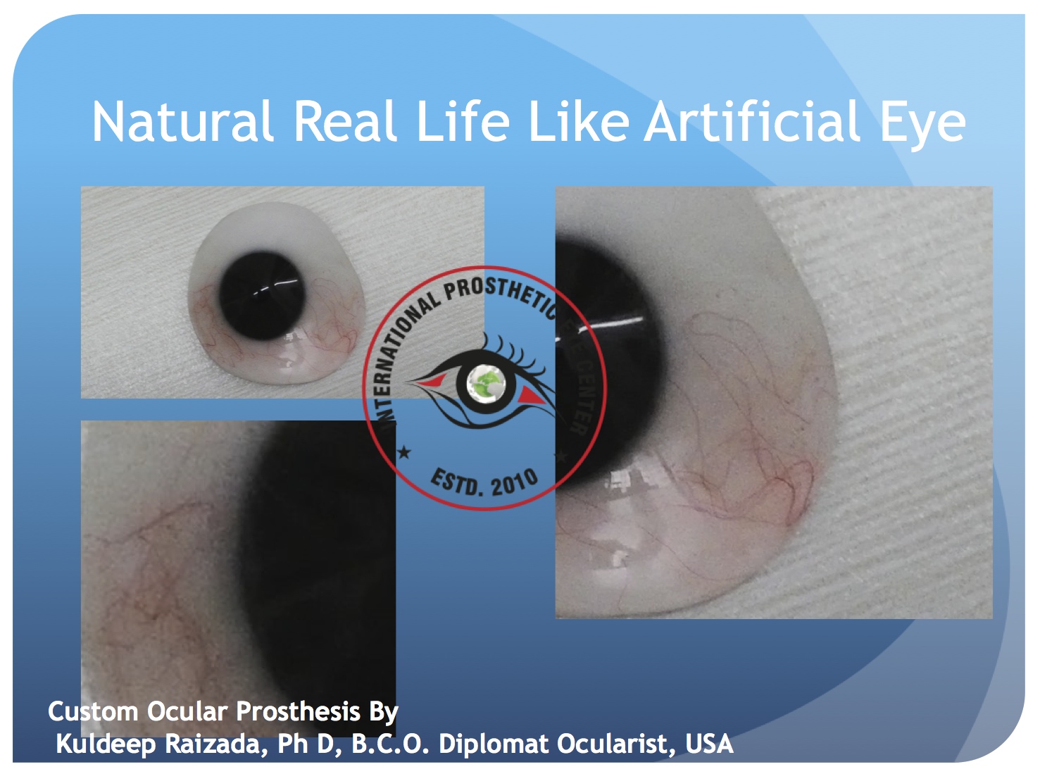
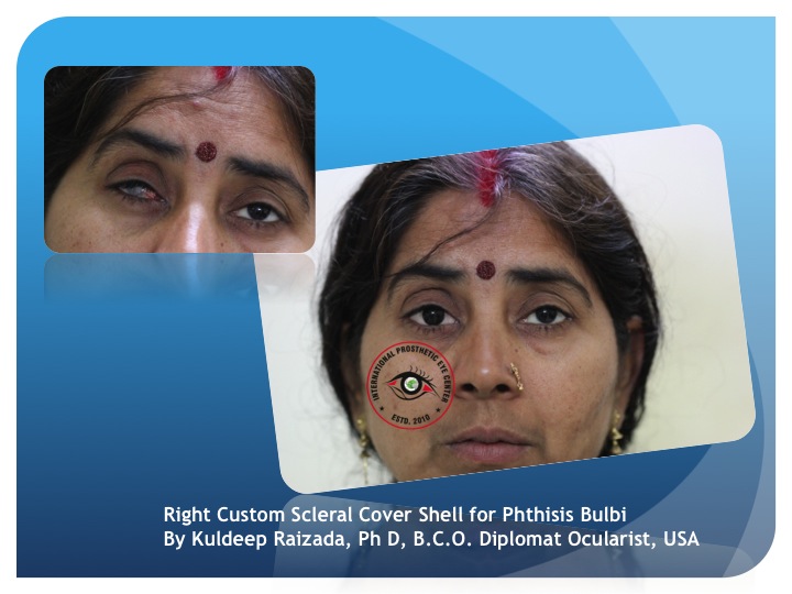
If the all treatment fails an ocular prosthesis is desired, many times it is is very hard to have very symmetrical eye prosthesis, at International prosthetic Eye Center we try to make as life like possible,
Reference:
1. Ahmad S, Osei-Bempong C, Dana R, Jurkunas U. The culture and transplantation of human limbal stem cells. Journal of cellular physiology 2010;225(1):15-9 doi: 10.1002/jcp.22251[published Online First].
2. Sangwan VS. Limbal stem cells in health and disease. Bioscience reports 2001;21(4):385-405
3. Strungaru MH, Mah D, Chan CC. Focal limbal stem cell deficiency in Turner syndrome: report of two patients and review of the literature. Cornea 2014;33(2):207-9 doi: 10.1097/ICO.0000000000000040[published Online First].
4. Skeens HM, Brooks BP, Holland EJ. Congenital aniridia variant: minimally abnormal irides with severe limbal stem cell deficiency. Ophthalmology 2011;118(7):1260-4 doi: 10.1016/j.ophtha.2010.11.021[published Online First].
5. Hatch KM, Dana R. The structure and function of the limbal stem cell and the disease states associated with limbal stem cell deficiency. International ophthalmology clinics 2009;49(1):43-52 doi: 10.1097/IIO.0b013e3181924e54[published Online First].
6. Di Iorio E, Kaye SB, Ponzin D, et al. Limbal stem cell deficiency and ocular phenotype in ectrodactyly-ectodermal dysplasia-clefting syndrome caused by p63 mutations. Ophthalmology 2012;119(1):74-83 doi: 10.1016/j.ophtha.2011.06.044[published Online First].
7. Messmer EM, Kenyon KR, Rittinger O, Janecke AR, Kampik A. Ocular manifestations of keratitis-ichthyosis-deafness (KID) syndrome. Ophthalmology 2005;112(2):e1-6 doi: 10.1016/j.ophtha.2004.07.034[published Online First].
8. Fernandes M, Sangwan VS, Vemuganti GK. Limbal stem cell deficiency and xeroderma pigmentosum: a case report. Eye 2004;18(7):741-3 doi: 10.1038/sj.eye.6700717[published Online First].
9. Lim P, Fuchsluger TA, Jurkunas UV. Limbal stem cell deficiency and corneal neovascularization. Seminars in ophthalmology 2009;24(3):139-48 doi: 10.1080/08820530902801478[published Online First].
10. Aslan D, Ozdek S, Camurdan O, Bideci A, Cinaz P. Dyskeratosis congenita with corneal limbal insufficiency. Pediatric blood & cancer 2009;53(1):95-7 doi: 10.1002/pbc.21960[published Online First].
11. Puangsricharern V, Tseng SC. Cytologic evidence of corneal diseases with limbal stem cell deficiency. Ophthalmology 1995;102(10):1476-85
12. Tsai RJ, Li LM, Chen JK. Reconstruction of damaged corneas by transplantation of autologous limbal epithelial cells. The New England journal of medicine 2000;343(2):86-93 doi: 10.1056/NEJM200007133430202[published Online First].
13. Meller D, Fuchsluger T, Pauklin M, Steuhl KP. Ocular surface reconstruction in graft-versus-host disease with HLA-identical living-related allogeneic cultivated limbal epithelium after hematopoietic stem cell transplantation from the same donor. Cornea 2009;28(2):233-6 doi: 10.1097/ICO.0b013e31818526a6[published Online First].
14. Sangwan VS, Jain V, Vemuganti GK, Murthy SI. Vernal keratoconjunctivitis with limbal stem cell deficiency. Cornea 2011;30(5):491-6
15. Uchino Y, Goto E, Takano Y, et al. Long-standing bullous keratopathy is associated with peripheral conjunctivalization and limbal deficiency. Ophthalmology 2006;113(7):1098-101 doi: 10.1016/j.ophtha.2006.01.034[published Online First].
16. Dua HS, Saini JS, Azuara-Blanco A, Gupta P. Limbal stem cell deficiency: concept, aetiology, clinical presentation, diagnosis and management. Indian journal of ophthalmology 2000;48(2):83-92
17. Dua HS, Azuara-Blanco A. Allo-limbal transplantation in patients with limbal stem cell deficiency. The British journal of ophthalmology 1999;83(4):414-9
18. Sridhar MS, Vemuganti GK, Bansal AK, Rao GN. Impression cytology-proven corneal stem cell deficiency in patients after surgeries involving the limbus. Cornea 2001;20(2):145-8
19. Ding X, Bishop RJ, Herzlich AA, Patel M, Chan CC. Limbal stem cell deficiency arising from systemic chemotherapy with hydroxycarbamide. Cornea 2009;28(2):221-3 doi: 10.1097/ICO.0b013e318183a3bd[published Online First].
20. Lichtinger A, Pe'er J, Frucht-Pery J, Solomon A. Limbal stem cell deficiency after topical mitomycin C therapy for primary acquired melanosis with atypia. Ophthalmology 2010;117(3):431-7 doi: 10.1016/j.ophtha.2009.07.032[published Online First].
21. Kim BY, Riaz KM, Bakhtiari P, et al. Medically reversible limbal stem cell disease: clinical features and management strategies. Ophthalmology 2014;121(10):2053-8 doi: 10.1016/j.ophtha.2014.04.025[published Online First].
22. Jeng BH, Halfpenny CP, Meisler DM, Stock EL. Management of focal limbal stem cell deficiency associated with soft contact lens wear. Cornea 2011;30(1):18-23 doi: 10.1097/ICO.0b013e3181e2d0f5[published Online First].
23. Chan CC, Holland EJ. Severe limbal stem cell deficiency from contact lens wear: patient clinical features. American journal of ophthalmology 2013;155(3):544-49 e2 doi: 10.1016/j.ajo.2012.09.013[published Online First].
24. Tseng SC. Staging of conjunctival squamous metaplasia by impression cytology. Ophthalmology 1985;92(6):728-33
25. Schermer A, Galvin S, Sun TT. Differentiation-related expression of a major 64K corneal keratin in vivo and in culture suggests limbal location of corneal epithelial stem cells. The Journal of cell biology 1986;103(1):49-62
26. Shapiro MS, Friend J, Thoft RA. Corneal re-epithelialization from the conjunctiva. Investigative ophthalmology & visual science 1981;21(1 Pt 1):135-42
27. Tseng SC. Concept and application of limbal stem cells. Eye 1989;3 ( Pt 2):141-57 doi: 10.1038/eye.1989.22[published Online First].
28. Gipson IK. The epithelial basement membrane zone of the limbus. Eye 1989;3 ( Pt 2):132-40 doi: 10.1038/eye.1989.21[published Online First].
29. Dua HS, Azuara-Blanco A. Limbal stem cells of the corneal epithelium. Survey of ophthalmology 2000;44(5):415-25
30. Osei-Bempong C, Figueiredo FC, Lako M. The limbal epithelium of the eye--a review of limbal stem cell biology, disease and treatment. BioEssays : news and reviews in molecular, cellular and developmental biology 2013;35(3):211-9 doi: 10.1002/bies.201200086[published Online First].
31. Ahmad S. Concise review: limbal stem cell deficiency, dysfunction, and distress. Stem cells translational medicine 2012;1(2):110-5 doi: 10.5966/sctm.2011-0037[published Online First].
32. Sandali O, Gaujoux T, Goldschmidt P, Ghoubay-Benallaoua D, Laroche L, Borderie VM. Infectious keratitis in severe limbal stem cell deficiency: characteristics and risk factors. Ocular immunology and inflammation 2012;20(3):182-9 doi: 10.3109/09273948.2012.672617[published Online First].
33. Huang AJ, Tseng SC. Corneal epithelial wound healing in the absence of limbal epithelium. Investigative ophthalmology & visual science 1991;32(1):96-105
34. Dua HS, Joseph A, Shanmuganathan VA, Jones RE. Stem cell differentiation and the effects of deficiency. Eye 2003;17(8):877-85 doi: 10.1038/sj.eye.6700573[published Online First].
35. Barbaro V, Ferrari S, Fasolo A, et al. Evaluation of ocular surface disorders: a new diagnostic tool based on impression cytology and confocal laser scanning microscopy. The British journal of ophthalmology 2010;94(7):926-32 doi: 10.1136/bjo.2009.164152[published Online First].
36. SCG. T. Application of Corneal Impression Cytology to Study Conjunctival Transdifferentiation Defect: Centro Grafico Editoriale, Univesita Degli Studi Parma, Parma, Italy,, 1988.
37. Deng SX, Sejpal KD, Tang Q, Aldave AJ, Lee OL, Yu F. Characterization of limbal stem cell deficiency by in vivo laser scanning confocal microscopy: a microstructural approach. Archives of ophthalmology 2012;130(4):440-5 doi: 10.1001/archophthalmol.2011.378[published Online First].
38. Dua HS. The conjunctiva in corneal epithelial wound healing. The British journal of ophthalmology 1998;82(12):1407-11
39. Poon AC, Geerling G, Dart JK, Fraenkel GE, Daniels JT. Autologous serum eyedrops for dry eyes and epithelial defects: clinical and in vitro toxicity studies. The British journal of ophthalmology 2001;85(10):1188-97
40. Rathi VM, Sudharman Mandathara P, Vaddavalli PK, Dumpati S, Chakrabarti T, Sangwan VS. Fluid-filled scleral contact lenses in vernal keratoconjunctivitis. Eye & contact lens 2012;38(3):203-6 doi: 10.1097/ICL.0b013e3182482eb5[published Online First].
41. Tseng SC, Prabhasawat P, Barton K, Gray T, Meller D. Amniotic membrane transplantation with or without limbal allografts for corneal surface reconstruction in patients with limbal stem cell deficiency. Archives of ophthalmology 1998;116(4):431-41
42. Sangwan VS, Matalia HP, Vemuganti GK, Rao GN. Amniotic membrane transplantation for reconstruction of corneal epithelial surface in cases of partial limbal stem cell deficiency. Indian journal of ophthalmology 2004;52(4):281-5
43. Fernandes M, Sangwan VS, Rao SK, et al. Limbal stem cell transplantation. Indian journal of ophthalmology 2004;52(1):5-22
44. Shimazaki J, Higa K, Morito F, et al. Factors influencing outcomes in cultivated limbal epithelial transplantation for chronic cicatricial ocular surface disorders. American journal of ophthalmology 2007;143(6):945-53 doi: 10.1016/j.ajo.2007.03.005[published Online First].
45. Pellegrini G, Traverso CE, Franzi AT, Zingirian M, Cancedda R, De Luca M. Long-term restoration of damaged corneal surfaces with autologous cultivated corneal epithelium. Lancet 1997;349(9057):990-3 doi: 10.1016/S0140-6736(96)11188-0[published Online First].
46. Sangwan VS, Basu S, Vemuganti GK, et al. Clinical outcomes of xeno-free autologous cultivated limbal epithelial transplantation: a 10-year study. The British journal of ophthalmology 2011;95(11):1525-9 doi: 10.1136/bjophthalmol-2011-300352[published Online First].
47. Johnen S, Wickert L, Meier M, Salz AK, Walter P, Thumann G. Presence of xenogenic mouse RNA in RPE and IPE cells cultured on mitotically inhibited 3T3 fibroblasts. Investigative ophthalmology & visual science 2011;52(5):2817-24 doi: 10.1167/iovs.10-6429[published Online First].
48. Sangwan VS, Basu S, MacNeil S, Balasubramanian D. Simple limbal epithelial transplantation (SLET): a novel surgical technique for the treatment of unilateral limbal stem cell deficiency. The British journal of ophthalmology 2012;96(7):931-4 doi: 10.1136/bjophthalmol-2011-301164[published Online First].
49. Basu S, Fernandez MM, Das S, Gaddipati S, Vemuganti GK, Sangwan VS. Clinical outcomes of xeno-free allogeneic cultivated limbal epithelial transplantation for bilateral limbal stem cell deficiency. The British journal of ophthalmology 2012;96(12):1504-9 doi: 10.1136/bjophthalmol-2012-301869[published Online First].
50. Brown KD, Low S, Mariappan I, et al. Plasma polymer-coated contact lenses for the culture and transfer of corneal epithelial cells in the treatment of limbal stem cell deficiency. Tissue engineering. Part A 2014;20(3-4):646-55 doi: 10.1089/ten.TEA.2013.0089[published Online First].
51. Sejpal K, Yu F, Aldave AJ. The Boston keratoprosthesis in the management of corneal limbal stem cell deficiency. Cornea 2011;30(11):1187-94 doi: 10.1097/ICO.0b013e3182114467[published Online First].
52. Espana EM, Di Pascuale M, Grueterich M, Solomon A, Tseng SC. Keratolimbal allograft in corneal reconstruction. Eye 2004;18(4):406-17 doi: 10.1038/sj.eye.6700670[published Online First].
53. Daya SM, Bell RW, Habib NE, Powell-Richards A, Dua HS. Clinical and pathologic findings in human keratolimbal allograft rejection. Cornea 2000;19(4):443-50
54. Liang L, Sheha H, Tseng SC. Long-term outcomes of keratolimbal allograft for total limbal stem cell deficiency using combined immunosuppressive agents and correction of ocular surface deficits. Archives of ophthalmology 2009;127(11):1428-34 doi: 10.1001/archophthalmol.2009.263[published Online First].
55. Cauchi PA, Ang GS, Azuara-Blanco A, Burr JM. A systematic literature review of surgical interventions for limbal stem cell deficiency in humans. American journal of ophthalmology 2008;146(2):251-59 doi: 10.1016/j.ajo.2008.03.018[published Online First].
56. Nishida K, Yamato M, Hayashida Y, et al. Corneal reconstruction with tissue-engineered cell sheets composed of autologous oral mucosal epithelium. The New England journal of medicine 2004;351(12):1187-96 doi: 10.1056/NEJMoa040455[published Online First].
57. Satake Y, Yamaguchi T, Hirayama M, et al. Ocular surface reconstruction by cultivated epithelial sheet transplantation. Cornea 2014;33 Suppl 11:S42-6 doi: 10.1097/ICO.0000000000000242[published Online First].
58. Kenyon KR, Tseng SC. Limbal autograft transplantation for ocular surface disorders. Ophthalmology 1989;96(5):709-22; discussion 22-3
59. Tan DT, Ficker LA, Buckley RJ. Limbal transplantation. Ophthalmology 1996;103(1):29-36
60. Baylis O, Figueiredo F, Henein C, Lako M, Ahmad S. 13 years of cultured limbal epithelial cell therapy: a review of the outcomes. Journal of cellular biochemistry 2011;112(4):993-1002 doi: 10.1002/jcb.23028[published Online First].
61. Shortt AJ, Secker GA, Notara MD, et al. Transplantation of ex vivo cultured limbal epithelial stem cells: a review of techniques and clinical results. Survey of ophthalmology 2007;52(5):483-502 doi: 10.1016/j.survophthal.2007.06.013[published Online First].
62. Di Iorio E, Ferrari S, Fasolo A, Bohm E, Ponzin D, Barbaro V. Techniques for culture and assessment of limbal stem cell grafts. The ocular surface 2010;8(3):146-53
63. Rama P, Matuska S, Paganoni G, Spinelli A, De Luca M, Pellegrini G. Limbal stem-cell therapy and long-term corneal regeneration. The New England journal of medicine 2010;363(2):147-55 doi: 10.1056/NEJMoa0905955[published Online First].
64. Basu S, Taneja M, Narayanan R, Senthil S, Sangwan VS. Short-term outcome of Boston Type 1 keratoprosthesis for bilateral limbal stem cell deficiency. Indian journal of ophthalmology 2012;60(2):151-3 doi: 10.4103/0301-4738.94060[published Online First].
Posted by Kuldeep Raizada. Posted In : Ophthalmology


 Kuldeep Raizada completed his basic optometry education at Gandhi Eye Hospital, Aligarh, and has his training at L V Prasad Eye Institute, Hyderabad. where he was also Founder and Head of the Department of Ocular Prosthesis services till 2009. He completed a second fellowship, in Anaplastolgy, at MD Anderson Cancer Centre, Houston. He has also been trained by the top most ocularist and anaplastologist in United States of America.
His clinical interests include ocular and facial prosthesis, particularly in pediatric patients. His research interests lie in newer advancement in development of new types of prosthesis, newer solution for ptosis corrective glasses.
Kuldeep Raizada, is Founder & Director of the International Prosthetic Eye Center since 2010, where he is practicing since 2010.
Kuldeep Raizada has been recognized by the American Society of Ocularist, USA and American Anaplastology Association,USA and by several other professional organizations, for his excellence in research and clinical practice.
Kuldeep Raizada, have completed all requirements by American Society of Ocularist, which is hard work of 14000 working hours as well extensive study for prosthetics, Hence awarded the Diplomate Ocularist from American Society of Ocularist, USA, 2012, Chicago, USA, which is the First ever received all over Asia Pacific & throughout Middle East so ever.
At present he is reviewer of several journals like Contact Lens & Anterior Eye, International Journal of Anaplastology, Oculoplasty & Reconstructive Surgery (OPRS) and Many others. He has published and presented world widely
